
Bausch & Lomb Microscope ISTX-8L: A Comprehensive Guide
This manual provides detailed instructions for the Bausch & Lomb ISTX-8L microscope, ensuring optimal performance and longevity. Explore its features and capabilities today!
The Bausch & Lomb ISTX-8L microscope represents a robust and reliable optical instrument, widely utilized in educational settings, clinical laboratories, and research facilities. This model is designed for versatility, supporting a range of microscopy techniques to observe diverse specimen types. Its construction prioritizes durability and ease of use, making it suitable for both novice and experienced users.
This guide serves as a comprehensive resource for understanding the ISTX-8L’s features, operation, and maintenance. Whether you are setting up the microscope for the first time, performing routine observations, or troubleshooting potential issues, this manual will provide clear and concise instructions. Proper understanding of its components and functions will unlock the full potential of this valuable scientific tool. We aim to empower you with the knowledge to achieve optimal imaging results consistently.
What’s Included in the Box?
Upon opening your Bausch & Lomb ISTX-8L microscope package, carefully verify the contents against the following list. You should find the ISTX-8L microscope head and base assembly, a set of four plan objective lenses (typically 4x, 10x, 40x, and 100x oil immersion), and a widefield eyepiece(s) – either binocular or monocular depending on the model purchased.
Additionally, the box contains a mechanical stage with specimen holder and controls, and an illumination system including a power supply and light bulb. You’ll also receive a dust cover to protect the microscope when not in use, and this comprehensive user manual. Small accessory items like immersion oil (for the 100x objective), and cleaning cloths are also included. If any items are missing or damaged, please contact Bausch & Lomb customer support immediately for assistance.
Safety Precautions
Before operating the Bausch & Lomb ISTX-8L microscope, please read and understand these crucial safety guidelines. Always ensure the microscope is placed on a stable, level surface to prevent accidental tipping. Never disassemble the microscope beyond the instructions provided in this manual; unauthorized repairs can be hazardous.
Exercise caution when handling glass slides and coverslips, as they can be fragile and sharp. When using the 100x oil immersion objective, only use approved immersion oil to avoid damaging the lens. Avoid looking directly at the high-intensity LED light source for extended periods. Always disconnect the power supply before cleaning or performing maintenance. Keep the microscope dry and protected from extreme temperatures and humidity. Follow proper electrical safety procedures and never operate the microscope with a damaged power cord.
Microscope Components & Their Functions
Explore the key parts of your ISTX-8L, including the optical system, head, nosepiece, stage, and illumination – each designed for precise observation and analysis.
Optical System Overview
The Bausch & Lomb ISTX-8L boasts a high-quality optical system engineered for exceptional clarity and resolution. This system meticulously gathers and focuses light, delivering sharp, detailed images of your specimens. It begins with the illumination source, passing through the condenser and diaphragm to control light intensity and contrast.
Light then travels through the objective lens – a critical component determining magnification and resolution – before reaching the eyepiece (ocular lens). The objective and eyepiece work in tandem to magnify the sample, allowing for detailed examination of cellular structures and other microscopic features.
Proper alignment of these components is crucial for optimal image quality. The ISTX-8L’s optical path is designed for minimal distortion, ensuring accurate representation of your sample. Understanding how each element contributes to the final image is key to maximizing the microscope’s potential and achieving reliable results in your observations.
Head (Binocular/Monocular) Details
The Bausch & Lomb ISTX-8L microscope head is available in both binocular and monocular configurations, catering to diverse user preferences and applications. The binocular head provides a more comfortable viewing experience, reducing eye strain during prolonged observation sessions through two eyepieces.
Interpupillary distance adjustment is a key feature of the binocular head, allowing users to customize the spacing between the eyepieces to match their individual eye separation for optimal viewing. Monocular heads, featuring a single eyepiece, offer a more compact and cost-effective option.
Both head types typically accommodate diopter adjustment on one eyepiece, compensating for differences in vision between the user’s eyes. Proper adjustment of the diopter ensures a clear, focused image for both eyes when using the binocular head. The head also allows for rotation, facilitating convenient viewing and sharing of observations.
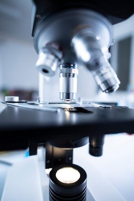
Nosepiece & Objective Lenses
The Bausch & Lomb ISTX-8L features a revolving nosepiece, typically accommodating four objective lenses, enabling users to quickly switch between different magnifications. These objective lenses are crucial for image formation, gathering light and magnifying the specimen. Common magnifications include 4x, 10x, 40x, and 100x (oil immersion).
Each objective lens is precisely engineered for optimal resolution and clarity at its designated magnification. The 100x objective requires immersion oil between the lens and the slide to enhance resolution and minimize light scattering. Proper handling and cleaning of objective lenses are essential to maintain image quality.
The nosepiece is designed for smooth and precise rotation, allowing for seamless transitions between objectives. Ensure the objective clicks securely into place before observation. Regularly inspect the lenses for dust or debris and clean them with lens paper and appropriate cleaning solutions.
Stage & Mechanical Controls
The Bausch & Lomb ISTX-8L microscope stage provides a stable platform for holding and manipulating slides. Typically, it features mechanical controls – coaxial knobs – for precise X-Y movement, allowing for smooth scanning of the specimen. These controls enable the user to systematically examine the entire slide area.
Many ISTX-8L models also include a stage clip to securely hold the slide in place. Some advanced stages may offer vernier scale measurements for accurate positioning and tracking of specific features. Proper adjustment of the stage height is crucial for optimal focusing and image clarity.
Regularly check the stage movement for smoothness and responsiveness. Avoid applying excessive force to the controls. Ensure the stage is clean and free of debris to prevent scratching of slides or objective lenses. Familiarize yourself with the stage controls for efficient and accurate specimen observation.
Illumination System
The Bausch & Lomb ISTX-8L’s illumination system is critical for producing clear, high-contrast images. It generally incorporates a halogen or LED light source, providing consistent and adjustable brightness. The intensity control allows users to optimize illumination based on the specimen and objective lens used.
A key component is the field diaphragm, which controls the diameter of the illuminated area, reducing glare and improving image contrast. The condenser, positioned beneath the stage, focuses the light onto the specimen. Adjusting the condenser height and aperture diaphragm is essential for achieving optimal resolution.
Regularly inspect the light source for proper functioning and replace bulbs as needed. Ensure the condenser and diaphragms are clean and free of dust. Proper illumination is fundamental to successful microscopy, enabling detailed observation of specimen structures.
Setting Up Your Microscope
Carefully unpack and position the ISTX-8L on a stable surface. Connect the power supply and familiarize yourself with the basic controls for optimal setup.
Powering On & Initial Adjustments
Begin by connecting the microscope to a grounded power outlet, ensuring the voltage selector switch matches your local supply. Upon powering on, the illumination system will activate. Allow a brief warm-up period for stable light output.
Start with the lowest power objective lens (typically 4x) in position. Adjust the interpupillary distance on the head to match your eye separation for comfortable binocular viewing. Focus initially using the coarse focus knob, then refine with the fine focus knob to achieve a sharp image.
The condenser should be in its lowest position, and the diaphragm partially closed. These initial adjustments provide a foundation for optimal image quality. Experiment with these controls later to enhance contrast and resolution. Remember to always start with low magnification for initial observation and gradually increase as needed.
Preparing Slides for Observation

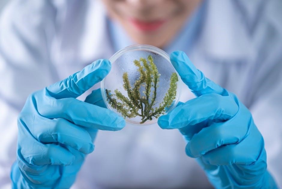
Proper slide preparation is crucial for clear observation. Begin with a clean glass slide and coverslip. For liquid samples, apply a small drop to the slide and gently lower the coverslip at an angle to avoid air bubbles. Solid samples may require mounting in a suitable medium.
If using a pre-made slide, ensure the specimen is centered on the stage. For wet mounts, a drop of immersion oil can be added to the coverslip to improve resolution, particularly at higher magnifications. Always use appropriate staining techniques to enhance visibility of cellular structures.
Secure the slide firmly with the stage clips. Before observation, carefully clean any excess mounting medium or oil from the slide’s surface. Proper preparation minimizes artifacts and maximizes image clarity, leading to accurate and reliable results during microscopic examination.
Focusing & Image Clarity
Achieving optimal focus is essential for detailed observation. Begin with the lowest power objective lens and utilize the coarse focus knob to bring the specimen into approximate focus. Then, switch to the fine focus knob for precise adjustments, ensuring a sharp and clear image.
Avoid over-correction, which can blur the image. Adjust the interpupillary distance and diopter settings on the head to compensate for individual eye differences. Proper alignment and adjustment contribute significantly to visual comfort and image quality.
For high-magnification objectives, use immersion oil to enhance resolution. Regularly check and adjust the condenser and diaphragm for optimal illumination and contrast. Consistent practice and careful observation will refine your focusing technique, yielding exceptional image clarity.
Using the Condenser & Diaphragm

The condenser focuses light onto the specimen, while the diaphragm controls the amount of light. Proper adjustment of both is crucial for optimal image contrast and resolution. Start with the condenser in its highest position and the diaphragm partially closed.
Adjust the condenser height to achieve even illumination across the field of view. Gradually open or close the diaphragm to control contrast; a smaller aperture increases contrast but reduces resolution, and vice versa. Experiment to find the ideal balance for your specimen.
For brightfield microscopy, a moderate diaphragm setting is generally best. Different techniques, like darkfield or phase contrast, require specific condenser and diaphragm settings. Refer to the relevant sections for detailed instructions. Consistent adjustment ensures clear, detailed observations.
Operating the ISTX-8L
Mastering the ISTX-8L unlocks detailed observation. Utilize its features for brightfield, darkfield, or phase contrast, achieving optimal results with each technique.
Brightfield Microscopy Techniques
Brightfield microscopy is the most common illumination method, ideal for observing stained specimens or those with inherent contrast. Begin by ensuring your slide is securely positioned on the stage and properly illuminated.
Adjust the condenser height and diaphragm aperture to optimize resolution and contrast; a lower aperture generally increases contrast but reduces resolution.
Start with the lowest power objective lens (e.g., 4x or 10x) to locate your specimen, then gradually increase magnification as needed.
Carefully utilize the coarse and fine focus knobs to achieve a sharp, clear image. Remember to center your specimen in the field of view before increasing magnification.
Proper specimen preparation, including appropriate staining techniques, is crucial for successful brightfield observation. Experiment with different staining methods to enhance visibility of specific cellular structures.
Always clean the objective lenses after use to maintain optimal image quality.
Darkfield Microscopy (If Applicable)
Darkfield microscopy reveals unstained specimens by illuminating them with light that does not directly enter the objective lens. This creates a bright background against which the specimen appears as a brightly lit object.
To achieve darkfield, adjust the condenser such that the darkfield stop is inserted into the light path, blocking direct light.
Proper alignment of the condenser and careful adjustment of the diaphragm are critical for optimal darkfield illumination.
Specimens should be relatively transparent and free of debris for best results.
Darkfield is particularly useful for observing live microorganisms, flagella, and other delicate structures that are difficult to visualize with brightfield microscopy.
Remember to minimize dust and scratches on slides, as these will scatter light and reduce image clarity.
Phase Contrast Microscopy (If Applicable)
Phase contrast microscopy enhances the contrast in transparent, unstained specimens, making internal structures visible without the need for staining. This technique exploits differences in refractive index within the sample.
The ISTX-8L, if equipped, utilizes phase contrast objectives and a specialized annular diaphragm within the condenser. Align the annular diaphragm with the objective’s phase ring for optimal results.
Adjust the phase contrast slider to maximize contrast and visibility of cellular details. Proper alignment is crucial; misaligned rings cause halos around structures.
Phase contrast is ideal for observing living cells and their dynamic processes, such as cell division and motility, without causing damage from staining.
Carefully clean phase objectives to avoid scratching the phase rings, which can degrade image quality.
Oil Immersion Techniques
Oil immersion microscopy increases resolution when viewing specimens at high magnification (typically 100x). It utilizes a special immersion oil with a refractive index similar to glass, minimizing light refraction.
Apply a small drop of immersion oil directly onto the coverslip over the specimen area. Carefully rotate the 100x objective into the oil, ensuring contact is made.
Avoid introducing air bubbles, as they will distort the image. Use only designated immersion oil; other oils can damage the objective lens.
Focus carefully using the fine adjustment knob. The image may appear blurry initially, but will sharpen with precise focusing.
After use, clean the objective lens immediately with lens paper and appropriate lens cleaning solution to prevent oil from drying and causing damage.

Maintenance & Troubleshooting
Regular cleaning and careful handling are crucial for maintaining optimal performance. This section details procedures for upkeep and resolving common operational issues.
Cleaning & Care of Optics
Maintaining pristine optics is paramount for clear, high-resolution imaging. Always begin by removing loose dust with a gentle blower or a soft brush designed specifically for lenses – avoid compressed air, as it can damage coatings.

For fingerprints or smudges, use lens cleaning paper moistened with lens cleaning fluid; apply gentle pressure in a circular motion, working from the center outwards. Never use abrasive cloths, tissues, or solvents like alcohol or xylene.
The oil immersion objective requires special attention. Immediately after use, remove immersion oil with lens paper moistened with xylene, followed by lens cleaning fluid. Inspect all optical surfaces regularly for scratches or damage. Proper storage, covered when not in use, will also extend the life of your lenses and ensure consistent image quality.
Bulb Replacement Procedures
Before replacing the illumination bulb, always disconnect the microscope from the power source to prevent electrical shock. Allow the bulb to cool completely before handling. Locate the bulb housing, typically accessible from the base of the microscope.
Carefully remove the housing cover, often secured with a screw or clip. Gently detach the old bulb from its connector – avoid touching the glass directly. Insert the new bulb, ensuring correct alignment with the connector. Use only the specified bulb type and wattage as indicated in the parts list.
Reattach the housing cover and secure it properly. Reconnect the microscope to the power source and test the illumination. If the bulb doesn’t light, double-check the connections and bulb compatibility. Dispose of the old bulb responsibly.
Common Issues & Solutions
If the image appears blurry, ensure proper focusing and clean the objective lenses with lens paper. Check slide preparation for air bubbles or debris. Dim illumination can often be resolved by adjusting the light intensity control or replacing the bulb.
For uneven illumination, adjust the condenser and diaphragm settings. If the microscope vibrates excessively, ensure it’s placed on a stable surface. Mechanical stage movement issues may require lubrication of the stage controls – consult a qualified technician.
If you encounter electrical problems, immediately disconnect the power. Never attempt to repair internal components yourself. Contact Bausch & Lomb support or a qualified microscope service provider for assistance. Always refer to the troubleshooting section in the full manual for detailed guidance.
Preventative Maintenance Schedule
To ensure optimal performance, implement a regular maintenance schedule. Weekly, clean all optical components – eyepieces, objectives, and condenser – with specialized lens paper. Dust the microscope body with a soft cloth to prevent buildup.
Monthly, inspect and tighten all screws and connections. Check the mechanical stage for smooth operation and lubricate if necessary. Quarterly, thoroughly clean the illumination system, including the bulb housing and condenser aperture.
Annually, consider a professional service check-up. This includes a complete optical alignment, mechanical inspection, and electrical safety assessment. Refer to the complete ISTX-8L manual for detailed instructions and recommended cleaning solutions. Consistent preventative care extends the microscope’s lifespan and maintains image quality.
Advanced Features & Capabilities
Unlock the ISTX-8L’s full potential! Explore digital imaging, polarization options, specialized filters, and precise calibration techniques for enhanced research outcomes.
Digital Imaging with the ISTX-8L
Capturing high-resolution images and videos with your Bausch & Lomb ISTX-8L microscope is now streamlined. This section details connecting compatible digital cameras – both dedicated microscopy cameras and DSLR adapters – to the phototube or trinocular head. We’ll cover software installation and configuration for image acquisition, processing, and annotation.
Proper camera selection is crucial; consider resolution, sensor size, and frame rate based on your application. Learn about adjusting exposure settings, white balance, and gain to optimize image quality. Explore techniques for image stacking to enhance depth of field and reduce noise.
Furthermore, we’ll discuss file formats (TIFF, JPEG, etc.) and their suitability for different purposes. Troubleshooting common issues like blurry images or software compatibility problems will also be addressed, ensuring seamless integration of digital imaging into your workflow. Detailed instructions and recommended software options are provided.
Polarization Microscopy (If Applicable)
If your Bausch & Lomb ISTX-8L is equipped for polarization microscopy, this section unlocks a powerful technique for analyzing birefringent materials. We’ll detail the function of the polarizer and analyzer, explaining how they interact with light to reveal structural details invisible under brightfield. Learn how to properly align these components for optimal contrast and image clarity.
Understanding retardation and optical path difference is key; we’ll cover these concepts and their impact on observed images. Explore different mounting techniques for samples, ensuring proper orientation for maximum birefringence visualization. This includes preparing thin sections of minerals, polymers, and biological tissues.
Furthermore, we’ll discuss applications like identifying crystalline structures, analyzing stress in materials, and observing muscle fiber organization; Troubleshooting common issues, such as uneven illumination or inaccurate readings, will also be addressed, maximizing the utility of this advanced microscopy method.
Using Filters for Specific Applications
The Bausch & Lomb ISTX-8L microscope supports a range of filters to enhance contrast, isolate specific wavelengths, and improve visualization for diverse samples. This section details filter placement and selection for optimal results. Explore neutral density filters for reducing light intensity when observing bright specimens, protecting your eyes and improving image quality.
We’ll cover blue filters for enhancing the visibility of stained cellular components, and green filters for improving contrast in unstained samples. Learn about excitation and emission filters used in fluorescence microscopy (if equipped), and their crucial role in visualizing fluorescently labeled specimens. Proper filter selection is paramount for accurate observation.
Troubleshooting filter-related issues, such as blurry images or color distortion, will also be addressed. A preventative maintenance schedule for cleaning filters will ensure long-term performance and prevent image degradation, maximizing the versatility of your ISTX-8L.
Calibration & Measurement
Accurate measurements are crucial in many microscopic applications, and the Bausch & Lomb ISTX-8L facilitates precise quantification. This section details the calibration process using a stage micrometer, establishing a known scale for accurate measurements of specimen dimensions. We’ll cover converting microscope units to real-world units (e.g., micrometers to millimeters).
Learn how to utilize the eyepiece reticle – a graduated scale within the eyepiece – for direct measurement of objects within the field of view. Understand the importance of consistent magnification during measurement to maintain accuracy. Explore methods for calculating the area of irregularly shaped objects using image analysis software (if applicable).
Troubleshooting common calibration errors, such as parallax or incorrect micrometer readings, will be addressed. A regular calibration schedule is recommended to ensure ongoing measurement precision, maximizing the scientific value of your ISTX-8L observations.